Next Steps

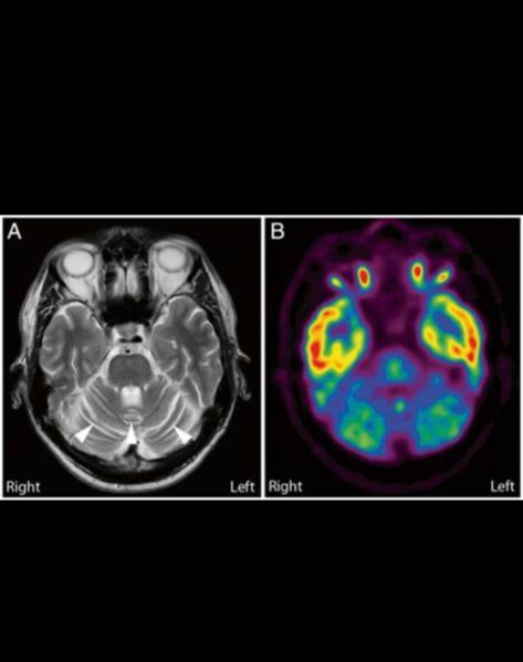
Yesterday I took Katie back to Vanderbilt for a follow-up. We had hoped her PET Scan/EEG would have given more comprehensive data but her condition and prior surgeries make the test difficult to pinpoint anything. We knew this may be the case but I was hoping we might get lucky. As suspected her right frontal lobe isn’t firing as it should. Her case was discussed at conference. Conclusion: More data needed. More testing

Next steps are for the pediatric neurosurgeon and the adult neurosurgeon need to connect after spring break but most likely we are looking at a stereo EEG. This is a more invasive EEG to better pinpoint where the seizures a generating from.

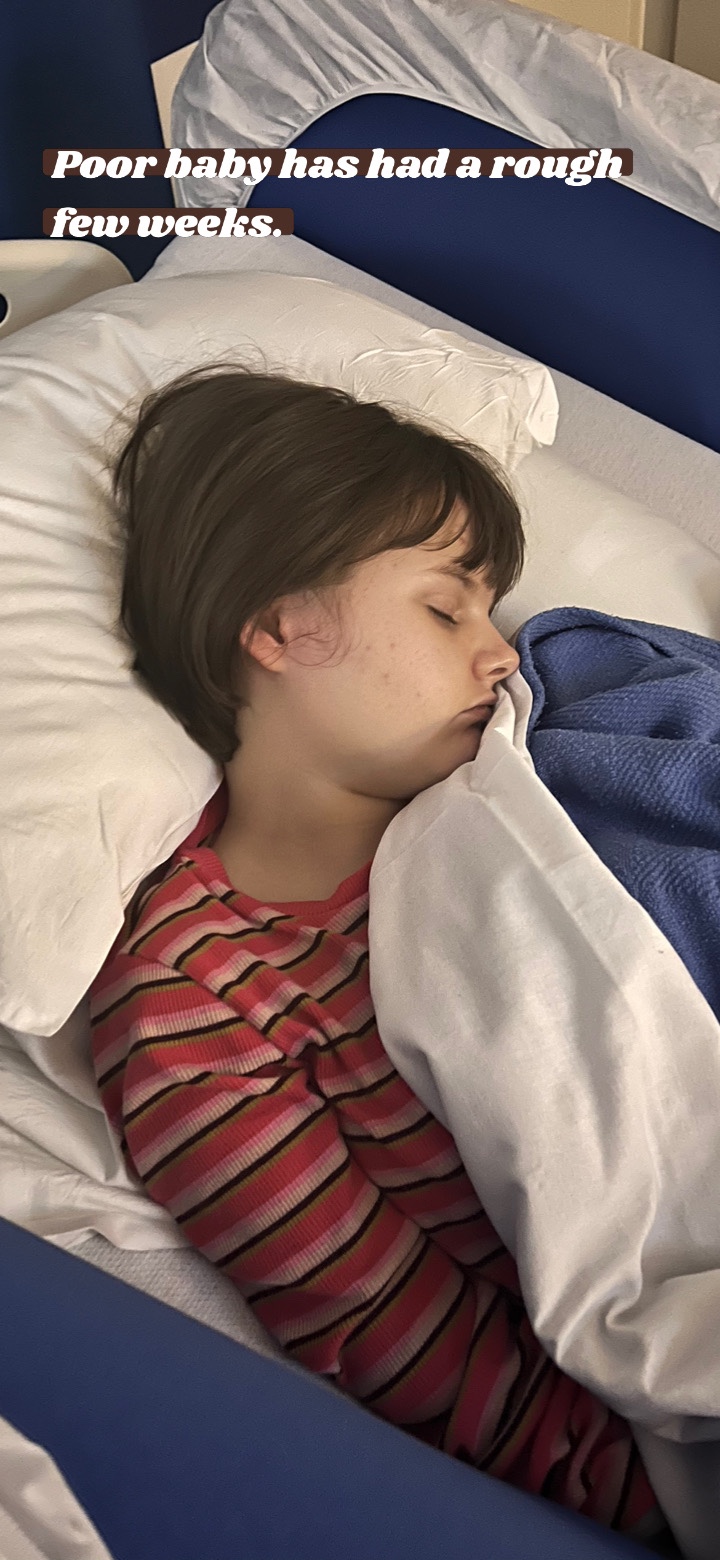
On a fantastic note, She has not had a seizure in 3 1/2 weeks. What a relief. We had gotten back to a place of 20-30 per day so this has been a nice break. A part of me wanted the neurologist to say, “Let’s just put things on hold for now.”But we know the big picture. We have seen time and time again over the past 15 years that she will have a honeymoon period after a procedure or med change. Sometimes they last a few months. Sometimes they last a few years. It is what it is. The RNS procedure is mostly likely going to be what changes her outlook longterm. We are pressing forward and hoping to get the next testing in another month or so.

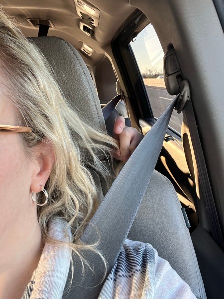
Today is day three of Spring Break. The boys are working so it’s just me and my shadow. We always have a love/annoy relationship going but after a day long rode trip…..she is on my last nerve. God love her. Bless her heart. Bless my heart. God help me. 5 hours in the van. All day. She napped on the way so lucky me. Headphones in. Crime podcast on. One the way back….not so lucky. She loves to lean forward and pull my hair or pull my seatbelt and try to choke me out. It’s real. All the while laughing hysterically. The other annoyance is she constantly wants to change out her DVD. She has a couple CD holders to organize them and she will flip through and perch the next movie on my shoulder to change it out. It’s like driving with a monkey.

So that’s the scoop. No fun Spring Break for us but hopefully we have lots of seizure free days coming. If you want to read more about her next text click the link below.

Stereoelectroencephalography (SEEG)

SEEG is the surgical implantation of electrodes into the brain in order to better localize the seizure focus. At UPMC, we use robotic assistance with ROSA® to accurately and efficiently place the electrodes for seizure mapping. Dr. Gonzalez was the first epilepsy surgeon in the US to offer SEEG and has performed over 1000 cases. He is also a pioneer of robotic-assisted neurosurgery, which improves accuracy and shortens surgery time.

https://www.neurosurgery.pitt.edu/centers/epilepsy/seeg

More Testing

This morning we drove to Vanderbilt children’s hospital. They hooked Katie up to an EEG and then did a metabolic PET scan of the brain. She did well. She always starts getting upset when someone comes in and puts gloves on. She got a quick EEG first and then they gave her an IV to administer the tracer. This is what shows at a cellular level how her brain is functioning.

After we headed to the cafeteria and got her the pizza that she’s been asking for all day. She wasn’t allowed to eat after midnight. I would have been seriously hangry and cranky myself.

I’m not sure how long it will take to get results back. I foresee the next level of testing will be several days in the hospital.

Thank you all for the comments, prayers and well wishes.

Here’s a bit of information about this test. Not an exciting read but I tried to just reference how the test is used for the brain; though this test is used for many other conditions.

What Is a Positron Emission Tomography (PET) Scan?

A PET scan is an imaging test that lets your doctor check for diseases in your body. The scan uses a special dye containing radioactive tracers. Certain organs and tissues absorb the tracer and help your doctor see how well your organs and tissues are working.

A positron emission tomography (PET) scan is an imaging test that allows your doctor to check for diseases in your body.

The scan uses a special dye containing radioactive tracers. These tracers are either swallowed, inhaled, or injected into a vein in your arm depending on what part of the body is being examined. Certain organs and tissues then absorb the tracer.

When detected by a PET scanner, the tracers help your doctor to see how well your organs and tissues are working.

The tracer will collect in areas of higher chemical activity, which is helpful because certain tissues of the body, and certain diseases, have a higher level of chemical activity. These areas of disease will show up as bright spots on the PET scan.

The scan can measure blood flow, oxygen use, how your body uses sugar, and much more.

A PET scan is typically an outpatient procedure, which means you can go about your day after the test is finished.

In the United States, around 2 million PET scans are performed each year, according to Berkley Lab.

Why is a PET scan performed?

Your doctor may order a PET scan to inspect your blood flow; your oxygen intake, or the metabolism of your organs and tissues. PET scans show problems at the cellular level, giving your doctor the best view of complex systemic diseases.

PET scans are most commonly used to detect:

• cancer

• heart problems

• brain disorders , including problems with the central nervous system.

Brain disorders

Glucose is the main fuel of the brain. During PET scans, tracers are “attached” to compounds such as glucose. By detecting radioactive glucose, the PET scan can show which areas of the brain are using glucose at the highest rates.

When a specialist interprets the scan, they can see how the brain is working and check for any irregularities.

How does the PET scan compare to other tests?

PET scans show metabolic changes occurring at the cellular level in an organ or tissue. This is important because diseases often begin at the cellular level. CT scans and MRIs cannot reveal problems at the cellular level.

PET scans can detect very early changes in your cells. CT scans and MRIs can only detect changes later, as a disease alters the structure of your organs or tissues

EEG’s, PET’s and MRI’s

Journal entry by Barbara Bradley — Oct 4, 2010

Another long day at Duke Hospital. We were there over three hours and met with the chief of neurology and another member of the epilepsy team. They reviewed her previous 24 hr EEG and got a more detailed understanding of her seizures. Dr. Mikati feels she would be a candidate for surgery as most children with her condition but he can’t proceed based on what we know. Only two seizures were captured so he wants her to have another EEG. Now that she is having so many, it shouldn’t be hard to get what they need. He wants her to have a PET scan. This test is over my head but the idea is to make sure the right side is functioning normally. The MRI shows it to be healthy but the PET scan will give us more information. He also wants to wean her off the depakote while raising her topamax dose over the next three weeks while we are waiting on getting her in for these other tests. Depakote can interfere with the blood platelets and cause bleeding (almost like aspirin). This is NOT what you want when undergoing brain surgery.

In the mean time, she is back to the way she was when we hospitalized her and having 2-3 seizures a night. We go back to living in the moment. On a good note, I had been in conversations with Johns Hopkins and shared her records with them. I heard back last week from one of their surgeons and he agreed she needed surgery and said Duke is very good and more than capable of handling the surgery. Their waiting list is even longer so his recommendation was to go with Duke if they are able to see her so quickly. This was a huge sigh of relief for us.